SMN1 Monoclonal Antibody
- Catalog No.:YM0588
- Applications:WB;IHC;IF;ELISA
- Reactivity:Human;Monkey
- Target:
- SMN1
- Gene Name:
- SMN1
- Protein Name:
- Survival motor neuron protein
- Human Gene Id:
- 6606
- Human Swiss Prot No:
- Q16637
- Mouse Swiss Prot No:
- P97801
- Immunogen:
- Purified recombinant fragment of human SMN1 expressed in E. Coli.
- Specificity:
- SMN1 Monoclonal Antibody detects endogenous levels of SMN1 protein.
- Formulation:
- Liquid in PBS containing 50% glycerol, 0.5% BSA and 0.02% sodium azide.
- Source:
- Monoclonal, Mouse
- Dilution:
- WB 1:500 - 1:2000. IHC 1:200 - 1:1000. IF 1:200 - 1:1000. ELISA: 1:10000. Not yet tested in other applications.
- Purification:
- Affinity purification
- Storage Stability:
- -15°C to -25°C/1 year(Do not lower than -25°C)
- Other Name:
- SMN1;SMN;SMNT;SMN2;SMNC;Survival motor neuron protein;Component of gems 1;Gemin-1
- Molecular Weight(Da):
- 32kD
- References:
- 1. J Med Genet. 2009 Sep;46(9):641-4.
2. RNA. 2009 Apr;15(4):515-23.
- Background:
- This gene is part of a 500 kb inverted duplication on chromosome 5q13. This duplicated region contains at least four genes and repetitive elements which make it prone to rearrangements and deletions. The repetitiveness and complexity of the sequence have also caused difficulty in determining the organization of this genomic region. The telomeric and centromeric copies of this gene are nearly identical and encode the same protein. However, mutations in this gene, the telomeric copy, are associated with spinal muscular atrophy; mutations in the centromeric copy do not lead to disease. The centromeric copy may be a modifier of disease caused by mutation in the telomeric copy. The critical sequence difference between the two genes is a single nucleotide in exon 7, which is thought to be an exon splice enhancer. Note that the nine exons of both the telomeric and centromeric copies are des
- Function:
- alternative products:Experimental confirmation may be lacking for some isoforms,disease:Defects in SMN1 are the cause of spinal muscular atrophy autosomal recessive type 1 (SMA1) [MIM:253300]. Spinal muscular atrophy refers to a group of neuromuscular disorders characterized by degeneration of the anterior horn cells of the spinal cord, leading to symmetrical muscle weakness and atrophy. Autosomal recessive forms are classified according to the age of onset, the maximum muscular activity achieved, and survivorship. The severity of the disease is mainly determined by the copy number of SMN2, a copy gene which predominantly produces exon 7-skipped transcripts and only low amount of full-length transcripts that encode for a protein identical to SMN1. Only about 4% of SMA patients bear one SMN1 copy with an intragenic mutation. SMA1 is a severe form, with onset before 6 months of age. SMA1 p
- Subcellular Location:
- Nucleus, gem . Nucleus, Cajal body . Cytoplasm . Cytoplasmic granule . Perikaryon . Cell projection, neuron projection . Cell projection, axon . Cytoplasm, myofibril, sarcomere, Z line . Colocalizes with actin and at the Z-line of skeletal muscle (By similarity). Under stress conditions colocalizes with RPP20/POP7 in punctuated cytoplasmic granules (PubMed:14715275). Colocalized and redistributed with ZPR1 from the cytoplasm to nuclear gems (Gemini of coiled bodies) and Cajal bodies (PubMed:11283611). Colocalizes with FMR1 in cytoplasmic granules in the soma and neurite cell processes (PubMed:18093976). .
- Expression:
- Expressed in a wide variety of tissues. Expressed at high levels in brain, kidney and liver, moderate levels in skeletal and cardiac muscle, and low levels in fibroblasts and lymphocytes. Also seen at high levels in spinal cord. Present in osteoclasts and mononuclear cells (at protein level).
- June 19-2018
- WESTERN IMMUNOBLOTTING PROTOCOL
- June 19-2018
- IMMUNOHISTOCHEMISTRY-PARAFFIN PROTOCOL
- June 19-2018
- IMMUNOFLUORESCENCE PROTOCOL
- September 08-2020
- FLOW-CYTOMEYRT-PROTOCOL
- May 20-2022
- Cell-Based ELISA│解您多样本WB检测之困扰
- July 13-2018
- CELL-BASED-ELISA-PROTOCOL-FOR-ACETYL-PROTEIN
- July 13-2018
- CELL-BASED-ELISA-PROTOCOL-FOR-PHOSPHO-PROTEIN
- July 13-2018
- Antibody-FAQs
- Products Images
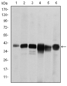
- Western Blot analysis using SMN1 Monoclonal Antibody against RAJI (1), Cos7 (2), Jurkat (3), K562 (4), HeLa (5) and HepG2 (6) cell lysate.
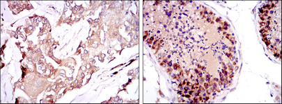
- Immunohistochemistry analysis of paraffin-embedded breast cancer tissues (left) and testis tissues (right) with DAB staining using SMN1 Monoclonal Antibody.
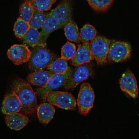
- Immunofluorescence analysis of HepG2 cells using SMN1 Monoclonal Antibody (green). Blue: DRAQ5 fluorescent DNA dye. Red: Actin filaments have been labeled with Alexa Fluor-555 phalloidin.