CHRDL1 Monoclonal Antibody
- Catalog No.:YM1022
- Applications:WB
- Reactivity:Human;Rat
- Target:
- CHRDL1
- Gene Name:
- CHRDL1
- Protein Name:
- Chordin-like protein 1
- Human Gene Id:
- 91851
- Human Swiss Prot No:
- Q9BU40
- Mouse Swiss Prot No:
- Q920C1
- Rat Gene Id:
- 363455
- Rat Swiss Prot No:
- Q76LD0
- Immunogen:
- Purified recombinant human CHRDL1 protein fragments expressed in E.coli.
- Specificity:
- CHRDL1 Monoclonal Antibody detects endogenous levels of CHRDL1 protein.
- Formulation:
- Liquid in PBS containing 50% glycerol, 0.5% BSA and 0.02% sodium azide.
- Source:
- Monoclonal, Mouse
- Dilution:
- WB 1:1000 - 1:2000. Not yet tested in other applications.
- Purification:
- Affinity purification
- Concentration:
- 1 mg/ml
- Storage Stability:
- -15°C to -25°C/1 year(Do not lower than -25°C)
- Other Name:
- CHRDL1;NRLN1;Chordin-like protein 1;Neuralin-1;Neurogenesin-1;Ventroptin
- Molecular Weight(Da):
- 52kD
- Background:
- This gene encodes an antagonist of bone morphogenetic protein 4. The encoded protein may play a role in topographic retinotectal projection and in the regulation of retinal angiogenesis in response to hypoxia. Alternatively spliced transcript variants encoding different isoforms have been described. [provided by RefSeq, Jan 2009],
- Function:
- function:Antagonizes the function of BMP4 by binding to it and preventing its interaction with receptors. Alters the fate commitment of neural stem cells from gliogenesis to neurogenesis. Contributes to neuronal differentiation of neural stem cells in the brain by preventing the adoption of a glial fate. May play a crucial role in dorsoventral axis formation. May play a role in embyonic bone formation (By similarity). May also play an important role in regulating retinal angiogenesis trough modulation of BMP4 actions in endothelial cells.,induction:By hypoxia in retinal pericytes.,similarity:Contains 3 VWFC domains.,tissue specificity:Expressed in retinal pericytes.,
- Subcellular Location:
- Secreted .
- Expression:
- Expressed in the developing cornea and in the eye anterior segment in addition to the retina. Differentially expressed in the fetal brain. There is high expression in cerebellum and neocortex. Expressed in retinal pericytes.
- June 19-2018
- WESTERN IMMUNOBLOTTING PROTOCOL
- June 19-2018
- IMMUNOHISTOCHEMISTRY-PARAFFIN PROTOCOL
- June 19-2018
- IMMUNOFLUORESCENCE PROTOCOL
- September 08-2020
- FLOW-CYTOMEYRT-PROTOCOL
- May 20-2022
- Cell-Based ELISA│解您多样本WB检测之困扰
- July 13-2018
- CELL-BASED-ELISA-PROTOCOL-FOR-ACETYL-PROTEIN
- July 13-2018
- CELL-BASED-ELISA-PROTOCOL-FOR-PHOSPHO-PROTEIN
- July 13-2018
- Antibody-FAQs
- Products Images
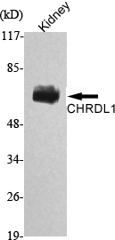
- Western Blot analysis using CHRDL1 Monoclonal Antibody against rat kidney lysate.